In the feline world, few anatomical features are as enigmatic as the nictitating membrane, commonly known as the third eyelid. This translucent or pale pink tissue, which sweeps horizontally across a cat’s eye, serves functions far beyond what meets the human eye. Often unnoticed by pet owners until it becomes prominent—a potential sign of illness—the third eyelid is a marvel of evolutionary adaptation. From acting as nature’s underwater goggles to serving as a silent health alarm, this structure is as functional as it is fascinating.
Unlike humans, cats possess this extra layer of ocular protection, a trait shared with many birds, reptiles, and some mammals. The membrane’s primary role is to shield the eye from debris, moisture loss, and UV radiation while maintaining visibility. When a cat blinks, the third eyelid slides across the cornea like a windshield wiper, distributing tears and removing dust. This mechanism is particularly vital for wild felines navigating dense foliage or engaging in high-speed chases where eye injuries could be catastrophic. Even domestic cats retain this evolutionary edge, though their urban lifestyles may not demand it as fiercely.
One of the most remarkable aspects of the nictitating membrane is its hydrodynamic prowess. For semi-aquatic species like the fishing cat or jaguar, the third eyelid acts as a natural diving mask. When submerged, it protects the cornea from irritation while allowing just enough light refraction for underwater vision. This adaptation hints at an ancestral link to environments where hunting required both terrestrial and aquatic agility. Though house cats may not swim often, the membrane’s residual functionality underscores how deeply ingrained these survival traits are.
Beyond its physical protections, the third eyelid is a barometer of feline health. Veterinarians often examine its prominence, color, and mobility to diagnose underlying conditions. A suddenly visible membrane—especially if bilateral—can signal dehydration, weight loss, or systemic illness like feline leukemia. Unilateral protrusion might indicate nerve damage or a localized infection. The membrane’s gland, responsible for tear production, is also prone to prolapse, leading to the cherry-like swelling known as "cherry eye." These clinical signs make the third eyelid an indispensable tool for early disease detection.
The membrane’s immunological role is equally critical. Its inner surface houses lymphoid tissue that secretes antibodies, creating a first line of defense against airborne pathogens. This feature is especially crucial for outdoor cats exposed to environmental microbes. However, this immune function can backfire when hyperactive responses lead to follicular conjunctivitis, causing discomfort and discharge. Such nuances highlight the delicate balance between protection and vulnerability in feline ocular health.
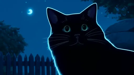
Cultural perceptions of the third eyelid have shifted over centuries. Ancient Egyptian art occasionally depicted cats with visible nictitating membranes, possibly interpreting them as a divine or mystical trait. Modern media, however, often misrepresents the membrane’s appearance—prolonged visibility in films or animations usually signifies a menacing or supernatural feline. This dramatization has inadvertently educated some viewers about its existence, though not always accurately.
For cat owners, understanding the third eyelid’s normal state versus abnormalities is crucial. A healthy membrane is typically inconspicuous, appearing briefly during sleep or sudden head movements. Persistent visibility, discoloration (yellowish in jaundice, red in inflammation), or sluggish movement warrants immediate veterinary attention. Routine checks during cuddle sessions can help owners establish baseline appearances, ensuring early intervention when deviations occur.
Research into the nictitating membrane continues to unveil its complexities. Recent studies explore its regenerative properties, with potential applications for human corneal repair. Other investigations focus on why certain breeds like Persians exhibit more prominent membranes, possibly linking to brachycephalic skull structures. Such findings not only advance veterinary medicine but also deepen our appreciation for this unassuming yet multifunctional tissue.
In essence, the cat’s third eyelid is a silent workhorse—simultaneously a guardian, a diagnostic tool, and an evolutionary relic. Its dual existence as both a practical asset and a health indicator encapsulates the intricate beauty of feline biology. For those who share their lives with cats, recognizing the stories hidden behind those fleeting glimpses of the nictitating membrane fosters a deeper connection to the creatures we cherish.
By /Jun 12, 2025
By /Jun 12, 2025
By /Jun 12, 2025
By /Jun 12, 2025
By /Jun 12, 2025
By /Jun 12, 2025

By /Jun 12, 2025
By /Jun 12, 2025

By /Jun 12, 2025

By /Jun 12, 2025
By /Jun 12, 2025
By /Jun 12, 2025
By /Jun 12, 2025

By /Jun 12, 2025
By /Jun 12, 2025
By /Jun 12, 2025
By /Jun 12, 2025